Dry Macular Degeneration Treatment | Dry AMD & Syfovre
Macular degeneration treatment depends on the stage of the disease and the type of macular degeneration present. Age-related macular degeneration (AMD) is the leading cause of vision loss in people over 50 in Australia and affects the macula, which is the central part of the retina responsible for detailed vision used for reading, recognising faces, and driving.
What Is the Macula?
The macula is a small central region of the retina that enables sharp, detailed vision. Because of this specialised function, the macula allows us to perform tasks that require precision.
For example, the macula helps with:
- reading
- recognising faces
- watching television
- driving
- using digital devices
To function properly, the macula relies on oxygen and nutrients from the choroid, a dense network of blood vessels beneath the retina.
Normally, Bruch’s membrane separates the retina from this vascular layer. This structure acts like a protective barrier and prevents fluid from leaking into the retina.
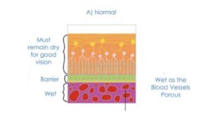
The macula is the central part of the retina responsible for detailed vision such as reading, recognising faces and watching television.
What Is Dry Macular Degeneration?
Dry macular degeneration develops when cells within the macula gradually become damaged over time.
In many cases, this process begins with the accumulation of drusen, which are small yellow deposits that form beneath the retina.
As these deposits increase, the retinal tissue may slowly thin. Consequently, central visual function can gradually decline.
Although small drusen may not immediately affect vision, larger deposits increase the risk of disease progression. Therefore, early detection plays an important role in monitoring the condition.
In clinical practice, eye care professionals often detect early macular degeneration during routine eye examinations. In addition, imaging techniques such as optical coherence tomography (OCT) help identify structural changes before noticeable symptoms develop.
Early detection allows doctors to monitor the disease carefully and consider treatment when appropriate.
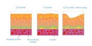
Diagram of dry macular degeneration showing drusen deposits and gradual degeneration of the macula
Drusen consist of cellular waste products that collect between the retina and the retinal pigment epithelium. While small drusen often do not affect vision, larger deposits increase the risk of progression to advanced disease. In many cases, clinicians detect early macular degeneration through a combination of your routine vision assessment and eye examinations, as well as OCT imaging before symptoms develop. Detecting the disease early allows the opportunity for earlier treatment commencement and therefore better long term visual outcomes.
Symptoms of Dry Macular Degeneration
Symptoms usually develop slowly and may be subtle in the early stages.
Common symptoms include:
- blurred central vision
- difficulty reading small print
- needing brighter lighting when reading
- distortion of straight lines
- reduced contrast sensitivity
Because these changes often occur gradually, the brain can compensate for small visual differences. As a result, many patients do not notice symptoms until the condition becomes more advanced.
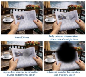
Progression of vision changes in age-related macular degeneration.
From left to right: normal vision, early distortion of straight lines, mid stage blurred and distorted central vision, and late stage loss of central vision.
Geographic Atrophy: An Advanced Stage of Dry AMD
In some patients, dry macular degeneration progresses to geographic atrophy (GA).
Geographic atrophy develops when areas of retinal cells gradually degenerate and stop functioning. As these cells disappear, well-defined patches of retinal loss become visible on retinal imaging.
Consequently, patients may notice:
- blurred or missing areas in central vision
- increasing difficulty recognising faces
- problems reading or seeing fine detail
- gradual worsening of central vision
Until recently, doctors could only provide monitoring and supportive care for this stage of the disease.
A New Treatment Option: Syfovre
More recently, new treatment options have emerged for patients with geographic atrophy.
Syfovre (pegcetacoplan) is a medication designed to slow the progression of geographic atrophy caused by dry macular degeneration.
The drug works by targeting part of the complement system, which plays an important role in retinal inflammation. By modifying this immune pathway, the treatment may reduce the rate of retinal cell degeneration.
Syfovre (pegcetacoplan) was approved after large phase 3 OAKS & DERBY clinical trials investigating treatment for advanced dry macular degeneration – geographic atrophy. The Clinical studies have shown that Syfovre may:
- slow the enlargement of geographic atrophy lesions
- reduce the rate of retinal degeneration in selected patients
However, it is important to understand that Syfovre does not restore vision that has already been lost. Instead, the goal of treatment is to slow further damage to the retina
How is Syfovre Treatment Is Given?
Syfovre is delivered as an intravitreal injection by your Ophthalmologist, meaning the medication is administered to the eye under anaesthetic.
Treatment is typically performed:
- in a procedure room at Eye and Laser Surgeons
- using local anaesthetic eye drops
- with sterile technique
Depending on the treatment plan, injections may be given every one to two months.
Dr Shanel Sharma will determine whether treatment is appropriate based on your retinal imaging, history and examination findings.
Who May Be Suitable for Syfovre?
Not all patients with macular degeneration are suitable candidates for this treatment.
Syfovre may be considered for patients with:
- geographic atrophy caused by dry macular degeneration
- evidence of disease progression on retinal imaging
- appropriate retinal anatomy on OCT scans
A detailed retinal assessment is required to determine suitability.
Monitoring Macular Degeneration
Regular monitoring plays a critical role in managing macular degeneration.
Ophthalmologists assess patients using several tests that evaluate both retinal structure and visual function.
These tests typically include:
- retinal examination
- optical coherence tomography (OCT)
- fundus photography
- fundus autofluorescence imaging
- visual function testing
Together, these investigations allow doctors to monitor disease progression and detect early changes in the macula.
Regular assessments are essential because they allow earlier detection of disease progression. As a result, treatment can begin at the most appropriate time.
In addition, your ophthalmologist may recommend monitoring your central vision at home using an Amsler grid. This simple grid test helps detect distortion of straight lines, which may signal worsening macular degeneration.
Images below show examples of how an Amsler grid may appear to patients with macular degeneration, including distortion of lines, blurred areas and central vision loss.
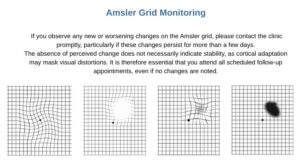
Examples of how an Amsler grid may appear to patients with macular degeneration, including distortion of lines, blurred areas and central vision loss.
When Should You Seek Assessment?
You should seek medical assessment promptly if you notice:
- distortion of straight lines
- blurred central vision
- increasing difficulty reading
- sudden changes in vision
These symptoms may indicate progression of macular degeneration. In some cases, they may also signal the development of wet age-related macular degeneration, which requires urgent treatment.
Therefore, if you notice any of these changes, you should arrange an eye examination within the next few days. To see Dr Sharma you can use this link to book now.
Macular Degeneration Care in Sydney
Macular degeneration requires accurate diagnosis and ongoing monitoring.
At Eye & Laser Surgeons in Sydney, patients undergo detailed retinal imaging to evaluate the stage of macular degeneration and discuss appropriate management options.
Dr Shanel Sharma and Dr Daya Sharma are specialist ophthalmologists who assess, diagnose and manage patients with macular degeneration and other retinal conditions.
Frequently Asked Questions
What is the difference between dry and wet macular degeneration?
In both dry and wet age-related macular degeneration (AMD), the disease process typically begins with the accumulation of waste material beneath the retina, forming deposits known as drusen within Bruch’s membrane.
In dry macular degeneration, retinal cells gradually degenerate over time. As a result, central vision slowly declines as the macula becomes less able to function normally.
In contrast, wet macular degeneration develops when the barrier function of Bruch’s membrane weakens. Consequently, choroidal blood vessels grow through this layer into the retina. Choroidal vessels are porous; therefore, they leak fluid or blood into the retina. This process damages the macula and can lead to rapid vision loss.
Diagram of dry macular degeneration showing drusen deposits and gradual degeneration of the macula
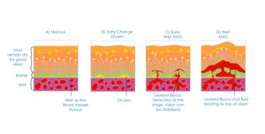
Illustration showing the progression of age-related macular degeneration from normal retina to drusen formation and wet AMD with abnormal blood vessel growth and leakage.
Can dry macular degeneration become wet?
Yes. Some patients with dry AMD may develop wet macular degeneration, which requires urgent treatment.
Does Syfovre cure macular degeneration?
No. Syfovre does not cure macular degeneration. Instead, the treatment aims to slow the progression of geographic atrophy, an advanced stage of dry AMD.
Is Syfovre suitable for everyone with dry AMD?
No. Doctors typically consider Syfovre only for patients with geographic atrophy, which represents an advanced stage of dry macular degeneration.
Further information about macular degeneration is available from the Macular Disease Foundation Australia.
Find out if you are suitable for vision correction
Not everyone is eligible for vision correction surgery.
Find out if you could benefit from this life changing surgery by taking the quick self-suitability quiz below:
Our most popular procedures
Take the first step toward clearer, healthier vision
Book an appointment to learn more about your eyes and the treatment options that may suit you
Take the first step toward clearer, healthier vision
Book an appointment to learn more about your eyes and the treatment options that may suit you

")